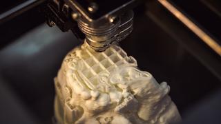
Additive manufacturing has been making waves within the medical industry for quite some time, but this week it has emerged that 3D printing has contributed towards the successful separation of conjoined twins.
Safa and Marwa Ullah were born fused at the skull, sharing a complex structure of blood vessels that made their separation an incredibly challenging task. Having already undergone two major operations, the twins were finally separated during the third procedure at Great Ormond Street Hospital in February 2019. Five months later, both twins are undergoing daily physiotherapy but are otherwise healthy.
Owing to the intricacies of these operations, it was crucial that surgeons were able to map out the individual anatomies of each twin ahead of time.
Owing to the intricacies of these operations - and the potentially tragic consequences resulting from errors - it was crucial that surgeons were able to map out the individual anatomies of each twin ahead of time. Additive manufacturing has been credited with making this possible, as surgeons were able to practice on 3D printed models that perfectly reflected the structures of the twins’ skulls and blood vessels. Together with the use of virtual reality, they were able to plan the best route for surgery, resulting in three successful operations that carried out the separation in distinct stages.
This is not the first time that additive manufacturing has been used in preparation for such a procedure. Whilst Safa and Marwa were born as craniopagus twins - that is, twins who are fused at the skull - the case of Knatalye and Adeline Mata presented surgeons with a different challenge. Born in Texas, fused from the chest all the way to the pelvis, the Mata twins were extensively conjoined, leaving doctors with no doubt as to the risks involved in surgery.
Taking a week to produce, the replica used multiple materials and could easily be assembled and disassembled to best aid planning efforts.
Fortunately, CT scans revealed that Knatalye and Adeline had separate hearts, although their livers were conjoined. This fusion of organs required precise planning from surgeons, meaning that once again, 3D printed models became a vital tool to negotiating a successful separation.
In this case, the model represented the twins’ skeletal and organ structures. Taking a week to produce, this replica used multiple materials and could easily be assembled and disassembled to best aid planning efforts. In addition, additive manufacturing was used to create a separate model of the liver anatomy complete with colour-coded blood vessels, allowing surgeons to identify the optimum separation site.
Following the successful separation procedure back in February 2015, Knatalye and Adeline Mata are now living normal lives.
Of course, conjoined twins are very rare, but additive manufacturing is allowing for extensive preparations within all types of major surgical operations. Brain surgery is one area in which there are very small error margins, with surgeons traditionally relying on MRI and CT scans alone to prepare for upcoming procedures of this nature.
One such case involved a woman with a tangled brain aneurysm. Theresa Flint was diagnosed with the condition after suffering recurring headaches, as well as problems with her vision. Although the aneurysm was too convoluted for regular surgery, failure to take action would have proven fatal. Therefore, surgeons needed to navigate the complexities of the aneurysm’s structure to decide upon the best course of action.
3D printing allows a higher degree of customisation when compared to conventional manufacturing.
After 3D printing a model of Theresa’s brain from a polymer that mimics human tissue, doctors took the time needed before surgery to come up with a solution. By planning thoroughly and ahead of time, surgeons were able to modify both their approach and the tools used to meet Theresa’s specific needs, resulting in a successful operation.
As a result, Theresa’s aneurysm has now been corrected without complications.
As well as assisting medical professionals prior to surgery, additive manufacturing is also making headway within the production of prosthetics and implants. 3D printing allows a higher degree of customisation when compared to conventional manufacturing, meaning that recipients can benefit from a prosthesis that replicates their own unique anatomy.
